Commercial Applications
- Properties, History and Challenges
- Overview
- Electric Power
- Transportation
- Medical Imaging and Diagnostics
- NMR for Medical and Materials Applications
- Industrial Processing
- High Energy Physics
- Wireless Communications
- Instrumentation, Sensors, Standards and Radar
- Large-Scale Computing
- Renewable Energy
- Cryogenics: An Enabling Technology
Applications in Medical Imaging and Diagnostics
MRI (Magnetic Resonance Imaging) has become the “gold standard” in diagnostic medical imaging, providing images of soft tissue not available from any other modality. It is not only safe and powerful, but thanks to superconducting magnets and their continued improvements, adds energy efficiency to its long list of benefits. In addition, advanced static and functional imaging techniques, using superconducting sensors, are emerging as complementary methods, enabling additional capabilities at lower cost. These include Ultra-Low-Field MRI, Magnetoencephalography (MEG) and Magnetocardiography (MCG). These advanced systems significantly improve the diagnostic tools available to healthcare providers and hold the promise of reducing lifetime healthcare costs.
Magnetic Resonance Imaging - MRI
Radiation-free Imaging.
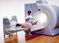 The introduction of MRI into the healthcare system has resulted in substantial benefits. MRI provides an enormous increase in diagnostic ability, clearly showing soft tissue features not visible using X-ray imaging or ultrasound. At the same time, MRI can often eliminate the need for harmful X-ray examinations. These advantages have greatly reduced the need for exploratory surgery. The availability of very precise diagnostic and location information is contributing to the reduction in the level of intervention that is required, reducing the length of hospital stays and the degree of discomfort suffered by patients.
The basic science of resonance imaging has been understood for many years. The nucleus of most atoms behaves like a small spinning magnet. When subjected to a magnetic field, the nuclear spin tries to align, but the spin means that instead it rotates around the field direction with a characteristic frequency proportional to the field strength. When a pulse of exactly the right radio frequency is applied, some of the energy of the pulse is absorbed by the nucleus, which can be measured as a signal that decays typically within several milliseconds. The timing of this signal decay, or relaxation, was discovered to depend critically on the chemical environment of the atom, and in particular was found to be different between healthy and diseased tissue in the human body. By rapidly switching on and off magnetic field gradients superimposed on the main field, it is possible to determine very accurate position information from these signals. The signals are processed by a computer to produce the now-familiar images from within the human body. Since the first crude MRI images were made in the 1970s, the industry has grown to a turnover of approximately $4B/year in new equipment sales. There are now well over 30,000 MRI systems installed worldwide, and the number is growing by 10% annually.
Advantages of Superconductivity
The heart of the MRI system is a magnet. The typical field values required for the latest generation of MRI cannot be achieved using conventional magnets. Just as importantly, high homogeneity and stability of the magnetic field are essential to achieve the resolution, precision and speed required for economical clinical imaging. The use of superconducting magnets provides a unique solution to these requirements. This trend will continue as field strength continues to progress from the current mainstream 1.5 T (Tesla) and 3 T systems to ever higher strengths in order to improve the clarity of the signal generated and increase the speed of image acquisition.
Expanding Applications of MRI
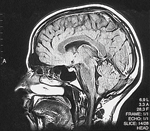 Functional Magnetic Resonance Imaging (FMRI), a rapidly growing extension of MRI techniques, uses a sequence of fast images to study dynamic changes, primarily blood flow rates. This has proved to be a powerful tool for imaging the activation of local regions in the brain. It is used to evaluate which areas of the brain are responsible for different functions, such as speaking, comprehension, moving fingers and toes and vision. An even newer technique, MRI guidance imaging, is used to assist physicians during surgery or ion beam therapy to plan the approach and more precisely locate, remove or radiate tumors. Another new technique, Magnetic Resonance Spectroscopy, is used on a limited basis for evaluating brain tumors, neurological diseases and epilepsy. Spectroscopy gives information on the chemical composition and metabolic activity of brain tissue. This information is used to assist in making diagnoses, monitoring changes and evaluating seizure activity. Further advances in the design of MRI systems are also enhancing the ability to utilize smaller, more specialized units dedicated to imaging of the body extremities such as arms and legs. The smaller foot-print of these units makes them more space, energy and resource efficient and allows for the higher throughput at a medical facility when combined with a whole body system for more complicated or extensive scans.
The Future of MRI
The number of MRI installations worldwide continues to grow at a rapid pace, providing ever more access to this powerful tool. MRI systems continue to advance in speed and resolution as the technologies of superconducting materials and superconducting magnets continue to advance. Exciting new methods that build on MRI are enabling new tools for both diagnosis and treatment of disease. It is clear that there are enormous potential benefits of continued support for R&D in superconducting materials and magnets.
Ultra-Low Field Magnetic Resonance Imaging (ULF-MRI)
Conventional Magnetic Resonance Imaging, discussed above, created a revolution in non-invasive imaging procedures, and the technique is used worldwide for many diagnoses. MRI is enabled by the high magnetic fields that only superconducting magnets can produce. Incremental improvements in the performance and cost of this established technology continue, but today researchers are also developing a complementary technique, Ultra-Low-Field MRI. In this new approach, instead of a high magnetic field from a superconducting magnet, a very low field - 10,000 times lower - is used. This low magnetic field is produced by simple, low cost magnets made with room temperature copper wire. To compensate for the loss of the high magnetic field, the extreme sensitivity of a superconducting detector is required. This detector, a “SQUID” (Superconducting Quantum Interference Device), enables the following benefits at low field:
- Significantly lower system cost, which could enable the new system to be much more widely available.
- Recent measurements on ex vivo prostate tissue demonstrate a significantly higher contrast between healthy and malignant tissue than at high fields. It is essential, however, to carry out studies to confirm that tumor imaging is viable in vivo. If ULF-MRI is successful in imaging cancer, it has a number of potential applications: diagnosing the severity of prostate cancer prior to biopsy, imaging of prostate cancer to guide biopsy, monitoring cancer progression during active surveillance or radiation therapy and imaging of other types of cancer, for example, brain and breast tumors.
- These two benefits combine to make ULF-MRI an important advance geared towards reducing the cost of healthcare on the one hand and enhancing the diagnostic ability of certain conditions on the other. The effort is slowly advancing from research to in vivo imaging; it holds also the promise of combining ULF-MRI with Magnetoencephalography (MEG). ULF-MRI is viewed by some as “greener” than high-field MRI in that it consumes vastly less electrical power, though helium usage is not as efficient.
Magnetoencephalography (MEG) and Magnetic Source Imaging (MSI)
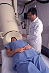 The same extreme sensitivity of SQUIDs that enables ULF-MRI has already enabled the development and
use of Magnetoencephalography (MEG), sometimes referred to as magnetic source imaging (MSI). In these systems, which are available commercially, an array of SQUID sensors detects magnetic signals from the brain in a totally non-invasive manner. Major successes include:[1]
- Pre-surgical mapping of brain tumors. By applying external stimuli (visual, audio, tactile), one can map out the function of the brain (which can be highly distorted by the presence of the tumor) prior to surgical removal of the tumor. With the aid of an MRI, this enables one to construct a 3D model of the brain and tumor.
- Showing the least invasive way of performing the surgery. This technique has been successful in reducing the incidence of collateral damage to the brain resulting from the surgical removal of the tumor.
- Location and pre-surgical mapping of the source of focal epilepsy. The focus is located using MSI. Pre-surgical mapping is conducted as for tumor surgery.
- Monitoring recovery from stroke or brain trauma (e.g. severe blow to the head as in football players, motorcycle accidents). MSI is used to monitor the response of the brain to standardized external stimuli (visual, audio, tactile) over a period of time to quantify the rate of recovery.
- completely non-invasive, requiring no electrode contact with the skin;
- provides wide-ranging information about the electrophysiological activity of the heart, including the detection of coronary artery disease; and
- signal strength depends on the distance between the heart and the detector, enabling the accurate measurement of the MCG of a fetus without saturating the detector with the signal from the mother’s heart (fetal-MCG or fMCG).
- [1] Clinical MEG system. Image courtesy 4-D Neuroimaging.
- [2] Non-invasive MCG system. Image courtesy CardioMag Imaging (CMI).
The use of MEG has been also extended to studies of unborn fetuses (fMEG). This technique has the potential to provide assessment of fetal neurological status and to assist physicians during high-risk pregnancies and diagnostics associated with infections, toxic insult, hypoxia, ischemia and hemorrhage. There are presently no other techniques for noninvasive assessment of fetal brain status.
The major challenge for the wider deployment of MEG systems is the initial cost of the system and the large database required to demonstrate excellent correlation of MSI with subsequent surgery. This effort involves system installation and data collection at research hospitals, an activity that is currently sparsely supported. The current initial diagnostic technique is low cost, Electroencephalography (EEG). The major advantage of MEG over EEG is that the former does not require any contact with the patient’s skin. In EEG, since electric currents travel the path of least resistance, moisture on the patient’s scalp and variations in skull thickness can distort the mapping of the epilepsy source.
Conversely, the magnetic field detected in MEG passes undistorted from the source to the SQUID detectors in the helmet worn by the patient. Since the interpretation of MSI inevitably requires an MR image, the combination of ULF-MRI with MSI into a single system would both reduce the cost of the combined procedures and improve their co-registration accuracy.
Magnetocardiography (MCG)
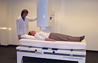 Sensitive SQUIDs are also the basis of functional
imaging of the heart in magnetocardiography (MCG
or MFI - heart magnetic field imaging) systems. MCG
systems detect, non-invasively and with
unprecedented accuracy, the net flows of cardiac
electric currents that drive the muscles in the heart.
In many clinical locations around the world, both
scientists and physicians are independently validating
the benefits of utilizing MCG for the detection and
diagnosis of many forms of heart disease, especially
cardiac ischemia and coronary artery disease.
Sensitivity for the detection of ischemia has been reported as high as 100% in recent
studies, and with such diagnostic accuracy it is not unreasonable to predict that MCG systems will find a home not only in hospitals, and especially emergency departments, but
also in outpatient imaging centers and cardiology clinics, where the rapid evaluation of
patients with suspicion of a life-threatening heart attack is absolutely critical to save lives.
Significant economic benefits can also be projected. Compared with electrocardiography
(EKG), MCG has a number of distinct advantages:[2]
While commercial systems do exist, the challenge for MCG, as in the case for MEG, is the development of a large enough database of clinical diagnostic correlations to convince insurers, such as Medicare, of the economic and healthcare benefits of MCG. Because it is radiation-free and risk-free, MCG can be used often during routine follow-up after an operation or during cardiac rehabilitation. The efficacy of a drug regimen can be tracked with MCG or even the recurrence of blockages after invasive treatment of a coronary artery. With the safety of a blood pressure reading and being equivalent to the diagnostic power of a magnetic resonance imaging procedure, MCG should be poised to revolutionize cardiac care.
Issues and Recommendations
The most pressing need in the quest to realize the full potential of these advances is the support for medical research aimed at correlating the data collected by ULF-MRI, MEG and MCG with actual clinical outcomes. This is critical in the quest to realize the full potential of these advances in systems and applications. Secondly, further support for superconductor development is needed to help provide the required performance, costs and availability to support the increasing array of applications that are looking to operate either with low or no helium present in the system. Helium availability and cost could represent one of the key challenges to further growth of these applications as currently they all rely on this commodity for cooling. While it is clear that future designs will look to reduce or eliminate helium entirely, currently the cost, performance and design constraints make this a very challenging choice.
